
Search Box (All Fields Optional)
Showing 1121 – 1140 out of 1141
Page 57 out of 58
| Thumbnails | Products | Price | Quantity | Action | |
|---|---|---|---|---|---|
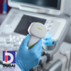
 | Ultrasound NT Scan – Accurate Nuchal Translucency Screening for Early Pregnancy The Ultrasound NT (Nuchal Translucency) Scan is a specialized prenatal screening test performed between 11 and 14 weeks of pregnancy to assess the risk of chromosomal abnormalities such as Down syndrome. It measures the fluid at the back of the baby’s neck using high-resolution ultrasound technology. Key features include non-invasive imaging, early detection of potential genetic conditions, and precise fetal development assessment. | د.إ650.00 | |||
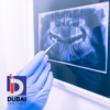
 | Comprehensive OPG Scan High-Quality Panoramic Dental Imaging The Comprehensive OPG Scan Panoramic Dental Imaging provides a detailed, full-mouth view of teeth, jawbones, and surrounding structures in a single, high-resolution scan. Utilizing advanced panoramic X-ray technology, it ensures precise diagnostics for dental conditions, including impacted teeth, bone abnormalities, and TMJ disorders. The non-invasive, quick, and painless procedure enhances patient comfort while delivering accurate, wide-angle imaging for efficient treatment planning. Ideal for dentists, orthodontists, and oral surgeons, this scan improves diagnostic accuracy, reduces radiation exposure, and streamlines dental assessments with clear, comprehensive visuals. | د.إ350.00 | |||
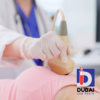
 | 3D/4D Pregnancy Ultrasound Scan A 3D/4D Pregnancy Ultrasound Scan provides expectant parents with a detailed, real-time view of their baby | د.إ950.00 | |||
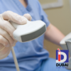
 | Renal Doppler Ultrasound Machine | Accurate Kidney Blood Flow Analysis Renal Doppler is a specialized ultrasound technique used to assess blood flow in the renal arteries and veins, aiding in the diagnosis of conditions like renal artery stenosis and hypertension. Key features include real-time color Doppler imaging, high-resolution spectral analysis, and advanced hemodynamic assessment. Its benefits include non-invasive evaluation, early detection of vascular abnormalities, and improved treatment planning. | د.إ950.00 | |||
 | Screening Mammography, Bilateral (2-View Film Study Of Each Breast) Screening Mammography, Bilateral (2-View Film Study of Each Breast) is a specialized X-ray imaging procedure designed for early breast cancer detection. This exam captures two high-resolution images of each breast, allowing radiologists to identify abnormalities such as tumors or microcalcifications before symptoms appear. Key benefits include early detection, which significantly improves treatment outcomes, and a non-invasive, quick procedure with minimal discomfort. Its unique selling points include advanced imaging technology for enhanced accuracy, routine use in preventive healthcare, and the ability to detect even small changes over time, making it a crucial tool in women’s health. | د.إ550.00 | |||
 | US Exam Abdominal (Renal/Aorta) Limited Ultrasound – Accurate Diagnostic Imaging The US EXAM ABDO (RENAL OR AORTA) LIMITED is a specialized ultrasound examination designed to assess the renal arteries, kidneys, or abdominal aorta for abnormalities such as blockages, aneurysms, or blood flow issues. This non-invasive, radiation-free diagnostic tool provides real-time imaging to aid in the early detection of vascular diseases, kidney disorders, or aortic aneurysms, ensuring timely medical intervention. Its high-resolution imaging enhances diagnostic accuracy, making it a safe, efficient, and cost-effective option for patients at risk of hypertension, kidney dysfunction, or aortic complications. | د.إ550.00 | |||
 | US Exam Abdo Back Wall – Ultrasound The US Exam Abdo Back Wall – Ultrasound is a specialized training tool designed to enhance ultrasound examination skills, particularly for assessing the abdominal back wall. It features realistic ultrasound imaging, high-fidelity anatomical structures, and durable, high-quality materials for repeated use. This model allows medical professionals and students to practice accurate probe positioning, image interpretation, and diagnostic techniques in a controlled environment. Its lifelike echogenic properties ensure a realistic scanning experience, making it an essential tool for improving proficiency in abdominal ultrasound assessments. Ideal for medical training programs, it enhances confidence, accuracy, and diagnostic efficiency in clinical practice. | د.إ550.00 | |||
 | US Exam Abdomen Complete – Ultrasound The US Exam Abdomen Complete – Ultrasound is a comprehensive diagnostic imaging procedure that evaluates abdominal organs, including the liver, gallbladder, pancreas, kidneys, spleen, and major blood vessels. It is a non-invasive, radiation-free test that provides real-time imaging to detect abnormalities such as tumors, cysts, gallstones, or organ enlargement. This ultrasound is safe, painless, and widely used for diagnosing conditions related to abdominal pain, liver disease, or kidney disorders. Its high-resolution imaging ensures accurate assessments, making it an essential tool for early detection and effective treatment planning. | د.إ550.00 | |||
 | US Exam Breast(s) – Ultrasound The US Exam Breast(s) – Ultrasound is a non-invasive imaging procedure designed to evaluate breast tissue for abnormalities, including cysts, tumors, and other concerns. It is particularly useful for women with dense breast tissue or those requiring further assessment after a mammogram. Key features include real-time imaging, radiation-free technology, and high-resolution visualization of soft tissues. Benefits include early detection of breast conditions, enhanced accuracy in differentiating between solid and fluid-filled masses, and suitability for all breast types. Its unique selling points are safe, painless screening, guidance for biopsies and interventions, and effectiveness in monitoring breast health over time. | د.إ650.00 | |||
 | US Exam of Head and Neck – Ultrasound The US Exam of Head and Neck – Ultrasound is a specialized imaging tool designed for precise evaluation of head and neck structures, including thyroid, lymph nodes, salivary glands, and vascular anatomy. It offers high-resolution imaging, real-time assessment, and non-invasive diagnostics, making it ideal for detecting abnormalities such as tumors, cysts, and vascular conditions. Key benefits include radiation-free imaging, enhanced diagnostic accuracy, and guidance for biopsies and interventions. Its portability, ease of use, and rapid results make it a valuable tool for clinicians in endocrinology, otolaryngology, and oncology. | د.إ550.00 | |||
 | USG ABDOMEN PELVIS KUB / ANY OTHER USG Non-Invasive Imaging: Uses high-frequency sound waves for real-time internal organ visualization.
Comprehensive Scanning: Covers abdomen, pelvis, kidneys, ureters, and bladder (KUB) for accurate diagnosis.
Radiation-Free: Safe for all patients, including pregnant women.
Doppler Capability: Assesses blood flow in organs and vessels. | د.إ550.00 | |||
 | د.إ550.00 | ||||
 | د.إ550.00 | ||||
 | د.إ550.00 | ||||
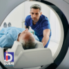 | Whole Body MRI Scan – Head, Chest & Abdomen | Comprehensive 3-Study Screening The Whole Body MRI Scan (3 studies: head, chest, abdomen) is a comprehensive, non-invasive diagnostic tool designed for early detection and monitoring of various health conditions. | د.إ3,750.00 | |||
 | Quick & Affordable X-Ray Single View Imaging Quick & Affordable X-Ray: Single View Imaging offers fast, cost-effective diagnostic imaging with a single-view X-ray. Designed for efficiency, it delivers high-quality images with minimal radiation exposure, making it ideal for routine screenings and preliminary assessments. Its affordability ensures accessibility for patients and healthcare providers, while rapid processing speeds enhance workflow efficiency. Perfect for urgent care, outpatient clinics, and budget-conscious facilities, this solution balances quality, speed, and cost-effectiveness for essential diagnostic needs. | د.إ150.00 | |||
 | Detailed X-Ray Two View Imaging | High-Resolution Diagnostic Scans Detailed X-Ray: Two View Imaging provides high-resolution, dual-angle X-ray scans for precise diagnostics. Key features include advanced imaging technology for enhanced clarity, rapid processing for quick results, and minimal radiation exposure for patient safety. Benefits include improved diagnostic accuracy, better treatment planning, and increased efficiency for healthcare providers. Its unique selling points are superior image detail, comprehensive two-view analysis for a more complete assessment, and optimized safety standards, making it an essential tool for accurate medical evaluations. | د.إ300.00 | |||
 | Comprehensive High-Quality X-Ray Three View Imaging for Accurate Diagnosis Comprehensive X-Ray: Three View Imaging provides high-quality, multi-angle diagnostic imaging for accurate assessments. Key features include three-view radiographic analysis (AP, lateral, and oblique), enhanced image clarity for precise diagnostics, and rapid processing for efficient patient care. Benefits include improved diagnostic accuracy, reduced need for additional imaging, and faster treatment planning. Its unique selling points are its comprehensive visualization, minimized patient exposure through optimized imaging techniques, and seamless integration with digital health systems for streamlined workflow. Ideal for medical professionals seeking reliable, in-depth radiographic evaluation. | د.إ400.00 | |||
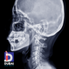 | Precise Cephalometric X-Ray Detailed Head & Jaw Analysis (CEPH) The Cephalometric X-Ray Detailed Head & Jaw Analysis is a specialized diagnostic tool used in orthodontics and maxillofacial treatment planning. It provides high-precision imaging of the skull, jaw, and dental structures, enabling accurate assessment of craniofacial relationships. Key features include high-resolution cephalometric imaging, precise anatomical measurements, and advanced software integration for in-depth analysis. Benefits include enhanced treatment planning, improved diagnostic accuracy, and better patient outcomes. Its unique selling points are automated landmark detection, customizable reporting, and seamless integration with orthodontic and surgical planning systems, making it an essential tool for orthodontists and maxillofacial specialists. | د.إ300.00 | |||
 | Cephalometric X-Ray & OPG Machine | High-Precision Dental Imaging System The Cephalometric X-Ray & OPG is an advanced dental imaging system designed for precise diagnosis and treatment planning in orthodontics and maxillofacial surgery. It combines Orthopantomogram (OPG) and Cephalometric X-ray capabilities, providing high-resolution, panoramic, and lateral skull images with minimal radiation exposure. Key features include automated image stitching, digital sensor technology, and adjustable positioning for patient comfort. Its fast scanning speed, superior image clarity, and user-friendly interface enhance diagnostic accuracy and workflow efficiency. Ideal for dental clinics and hospitals, this system ensures comprehensive craniofacial assessment, improved treatment outcomes, and seamless integration with digital practice management software. | د.إ400.00 |